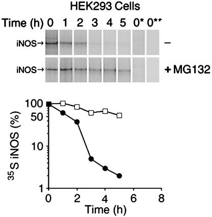
Fig. 1.
Determination of human iNOS half-life. HEK 293 cells, stably expressing human iNOS, were pulsed with [35S]methionine/cysteine for 1 h and chased with unlabeled media at various time points in the presence or absence of 10 μM proteasome inhibitor MG132. iNOS was immunoprecipitated with anti-iNOS antibody. To verify the stringency of immunoprecipitation, in some samples the immunoprecipitating antibody was omitted (lane 0*) or HEK 293 cells that do not express iNOS were used (lane 0**). Eluted proteins were separated by SDS/PAGE, and 35S-labeled iNOS was detected by PhosphorImager. (Lower) iNOS half-life was calculated, for cells cultured in the absence (filled circles) or the presence of MG132 (open squares), as the time needed for decay of 50% of 35S-labeled iNOS. The half-life of human iNOS in HEK 293 cells was 2.0 ± 0.1 h and was prolonged to 6.5 ± 1.2 h in cells treated with the proteasomal inhibitor MG132 (average ± SD, n = 3).

