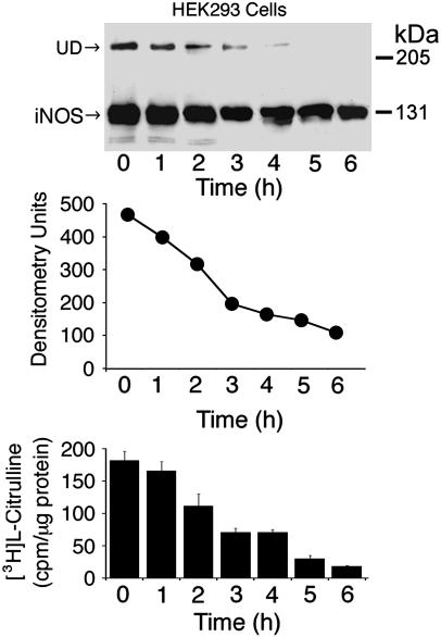
Fig. 3.
Time course of the effect of iNOS dimerization inhibitor, BBS-1. HEK 293 cells stably expressing human iNOS were incubated for the indicated time periods in the presence of 1 μM BBS-1. A representative Western blot of cells lysates (50 μg per lane), using an anti-iNOS antibody, is shown (Top). UD, presence of UD of iNOS. Densitometry analysis of bands representing iNOS on the Western blot is shown (Middle). iNOS activity was evaluated in cell lysates by the conversion of [3H]arginine to [3H]citrulline (Bottom) (means ± SD, n = 3).

