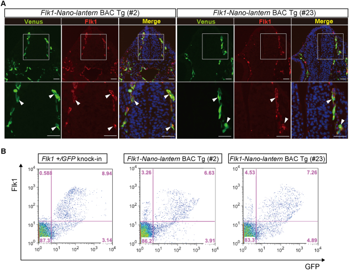
Figure 3. Faithful expression of Nano-lantern driven by the Flk1 regulatory sequence in vascular ECs.
Venus expression in Flk1-Nano-lantern BAC Tg embryos at 9.5 dpc. (A) Co-expression of Venus and the endogenous Flk1 protein. Transverse sections posterior to the heart of Flk1-Nano-lantern BAC Tg embryos (#2 and #23) were subjected to immunohistochemical analysis with anti-GFP and anti-Flk1 antibodies to further compare the distribution of their expression. Arrowheads indicate ECs with co-expression of Venus and Flk1. Scale bars: 50 μm. (B) Flow cytometric analysis of Flk1-Nano-lantern BAC Tg embryos at 9.5 dpc (#2 and #23). Single cells prepared from Flk1-Nano-lantern BAC Tg embryos was incubated with PE-labelled anti-Flk1 antibody and flow cytometry was performed as described in Materials and Methods.

