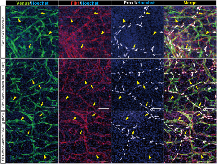
Figure 4. Expression of Flk1-Nano-lantern in lymphatic ECs.
Immunohistochemical analysis of the back skin of Flk1-Nano-lantern BAC Tg mice with anti-GFP, -Flk1 and –Prox1 antibodies. Arrows and arrowheads indicate Prox1-positive lymphatic ECs and Prox1-negative vascular ECs, respectively. Scale bars: 50 μm

