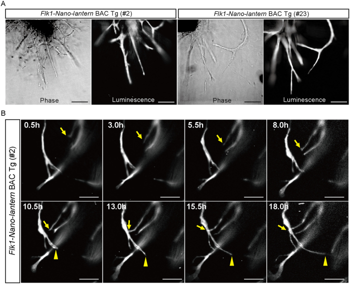
Figure 6. Luminescence imaging of ECs in aortic ECs of Flk1-Nano-lantern BAC Tg mice.
(A) Detection of luminescence in endothelial cells in the aortic rings from Flk1+/GFP mice and Flk1-Nano-lantern BAC Tg mice. The aortic rings were isolated from Flk1+/GFP mice and Flk1-Nano-lantern BAC Tg mice (#2 and #23) and cultured in the presence of VEGF-A. Luminescence was detected after the addition of coelenterazine-h to the media. Scale bars: 100 μm. (B) Time-lapse luminescence imaging of sprouting ECs. Luminescence was detected every 30 min and recorded over an 18 hr period. Arrows and arrowheads indicate sprouting ECs. Scale bars: 10 μm.

