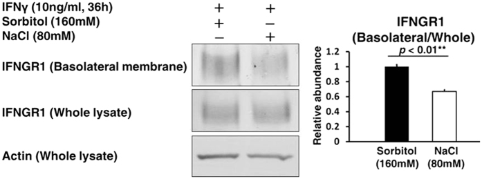
Figure 5. A trafficking defect of IFNGR1 to the basolateral membrane in HK2 cells exposed to a high salt concentration.
The cells were treated by IFNγ (10 ng/mL) for 36 h after a 2 h exposure of culture medium supplemented with either NaCl (80 mM) or Sorbitol (160 mM, as an osmotic control). Biotinylation assay was performed to evaluate the protein abundance of IFNGR1 in the basolateral membrane of HK2 cells. Left, Representative immunoblotting performed to evaluate the protein abundance of IFNGR1; Right, Densitometry analysis of the immunoblotting of the protein abundance of IFNGR1 in the basolateral membrane (n = 3). Full-length western blot images are presented in Supplementary Figure S4. Although the protein abundance of IFNGR1 in whole cell lysate was not markedly altered by a high salt concentration, the protein abundance of IFNGR1 in the basolateral membrane significantly decreased.

