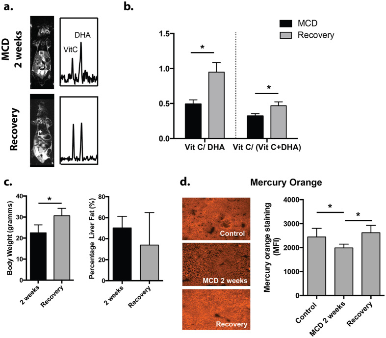
Figure 4. Study of recovery phase animals (MCDr group) by HP DHA.
(a) Representative 13C spectra obtained from the MCD group are compared to those obtained from mice first fed the MCD diet, then normal chow for 1 week. The HP DHA to VitC resonance ratios in the recovery group appear similar to those obtained for controls. (b) Bar graphs obtained for MCDr mice show significant increases in VitC/VitC + DHA and VitC/DHA ratios (p < 0.05) following return to a normal diet, which were not significantly different from baseline mice. (c) Although body weights in MCDr mice returned to normal, there was still significant hepatic steatosis (30%) seen in these animals, 2-fold that seen in the control group. (d) Mercury Orange staining showing decreased thiol content in MCD mice. Significant normalization was seen in the recovery group.

