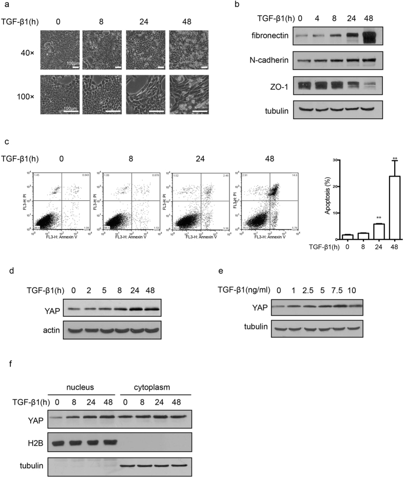
Figure 1. Increased YAP levels in TGF-β1-induced apoptosis and EMT.
(a) Cell morphological changes were detected in NMuMG cells after TGF-β1 (10 ng/mL) treatment for the indicated time. (b) The effect of TGF-β1 (10 ng/mL, 48 h) on the protein level of EMT markers was detected by immune blotting (Epithelial marker: ZO-1. Mesenchymal markers: fibronectin and N-cadherin). (c) The effect of TGF-β1 (10 ng/mL) on apoptosis was detected by Annexin V/PI double staining and analyzed by flow cytometry. The representative images (left) and statistical data (right) were shown. The data are the means ± SD of three independent experiments. (d) Time-dependent effect of increased YAP expression level was detected by immunoblotting after TGF-β1 (10 ng/mL) treatment in NMuMG cells. (e) The dosage-effect of TGF-β1 on the expression level of YAP. Cells were treated with different concentration of TGF-β1 for 48 h. (f) The cytoplasmic and nuclear YAP levels were examined after TGF-β1 (10 ng/mL) treatment for the indicated time.

