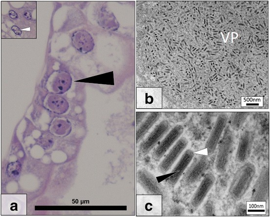
Fig. 2.

Gammarus roeselii Bacilliform Virus (GrBV) histopathology and ultrastructure. a Several virally infected, hypertrophic, nuclei (black arrow) in the hepatopancreas. Inset at the same magnification details a cluster of uninfected nuclei (white arrow). b Electron micrograph detailing a growing viroplasm (VP) in a nucleus of the hepatopancreas. c High magnification image of the bacilliform virus present with electron dense core (black arrow) and membrane (white arrow) in a paracrystalline array within a heavily infected cell nucleus. Scale-bars: a, 50 μm; b, 500 nm; c, 100 nm
