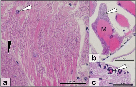
Fig. 3.

Cucumispora roeselii n. sp. histopathology. a Microsporidian spores (black arrow) can be seen throughout the musculature in heavy infections. Muscle nuclei (white arrow) can be seen amongst parasite spores. b Early stage microsporidian infected muscle blocks (M) demonstrate initial sarcolemma infection (white arrow). c Immune reactions (white arrow) towards microsporidian infection. Scale-bars: 50 μm
