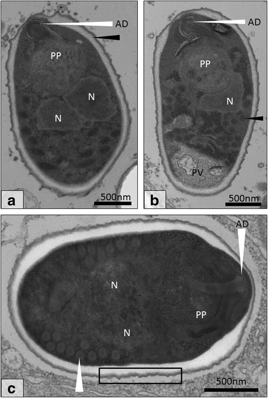
Fig. 5.

Final development stages of Cucumispora roeselii n. sp. a Diplokaryotic sporoblast (N) with anchoring disk (AD), polaroplast (PP) and thickened endospore (black arrow). b A second sporoblast displaying a clear polar vacuole (PV) and polar filament with rings of varying electron density (black arrow). c The final diplokaryotic (N) spore with bilaminar polaroplast (PP), anchoring disk (AD) and polar filament (9–10 turns; white arrow). The spore wall thins at the anchoring disk (AD) whilst being thickest at the periphery of the anchoring disk. Note the ‘thorned’ spore exterior (black rectangle). Scale-bars: 500 nm
