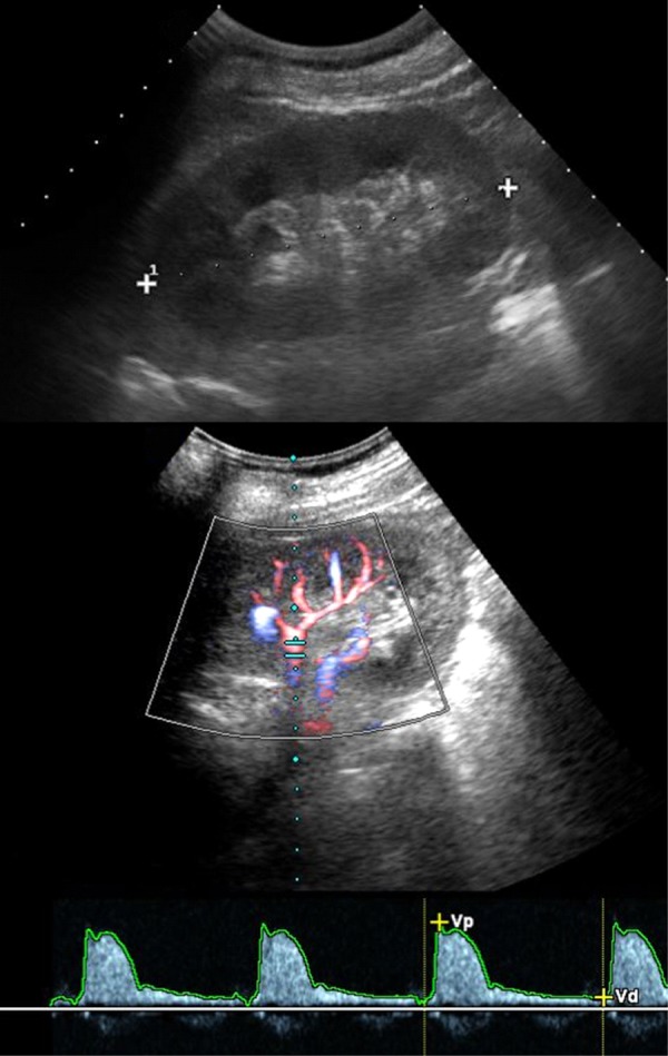
Figure 4. Examples of diabetic nephropathy.

The figure shows ultrasound images of a 50-year old type 1 diabetic patient with severe renal failure. Ultrasonography (upper image) shows normal appearance of the left kidney with regular corticomedullary differentiation and size. Color-Doppler sonography and Doppler spectrum of an intrarenal artery demonstrate a normal waveform and a normal early systolic compliance peak without abnormal findings.
