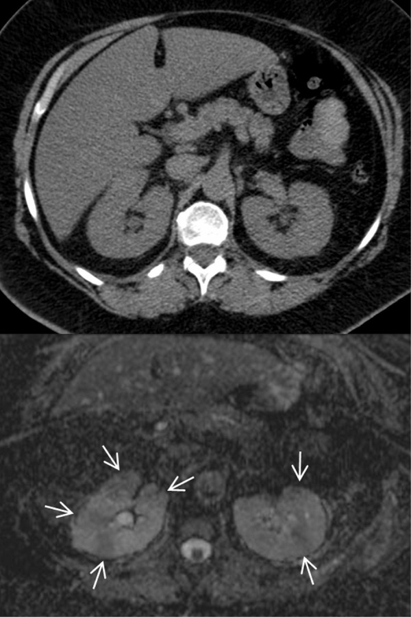
Figure 7. Diffusion-weighted MRI in a 38 years old patient with type 1 diabetes and acute bacterial pyelonephritis in the left kidney.

Native diffusion-weighted images obtained at b values of 0 and 150 sec/mm2 show some cortical wedge-shaped areas of faint hyper-intensity that are well detectable. The areas persist at the higher b value of 700 sec/mm2 (arrows) suggesting reduced diffusion due to inflammatory edema and acute pyelonephritis. This finding is confirmed by the apparent diffusion coefficient map obtained from b values of 150 and 700 sec/mm2, a procedure carried out to exclude vascular components detected by the MRI signal, which shows areas of low signal intensity (arrows; mean ADC value of 1.25 x 10⁻³mm2s-1) and healthy tissue for comparison reasons to see inflammatory parenchymal changes.
