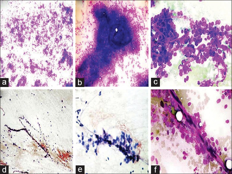
Figure 1.

(a) Hypercellular smears displaying monomorphic round cells, (MGG ×40). (b) Smears displaying clumps of opaque myxoid material obscuring round cells. A plexiform capillary is still discernible, (MGG ×100). (c) Round monomorphic cells with indistinct-to-distinct nucleoli and fine thin capillaries in between, (MGG ×400). (d) Smears showing thin plexiform capillaries along with associated neoplastic round cell, (Pap stain ×40). (e, f) Higher magnification displaying thin capillaries with adherence of round neoplastic cells (ANAC), (e: Pap stain ×100; f : MGG × 400)
