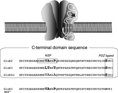
Fig. 2.
Sequence alignment of GluR2, GluR3, and GluR4c, shown for rat (≥98% identity to mouse homologs). Bold residues denote differences between GluR2 and GluR3 in the NSF-binding region. GluR3 NSF+ mutation = GluR3 L853V/T854A/T857P. Ser-880, located within the GluR2 PDZ ligand, as well as corresponding serines in other subunits, are also shown in bold.

