Figure 2.
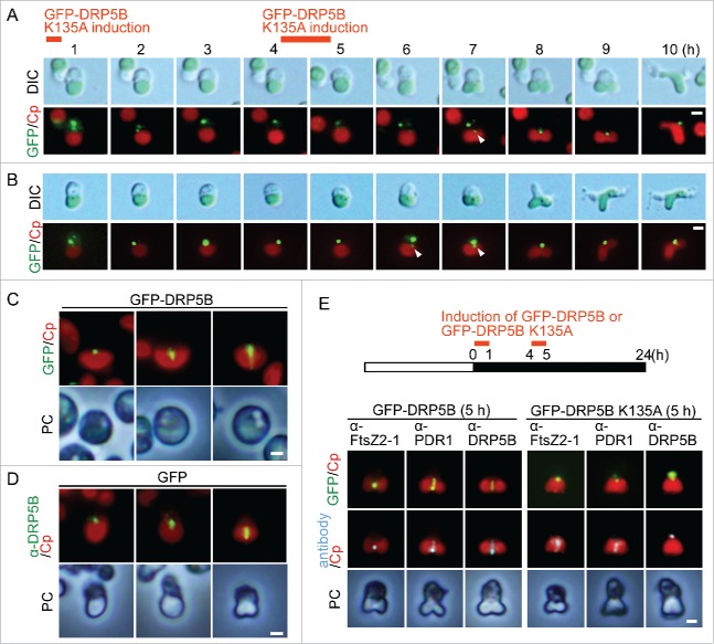
DRP5B ring formation and effect of GFP-DRP5B K135A expression before the onset of chloroplast division on the localization of chloroplast division proteins. (A, B) GFP-DRP5B K135A was expressed before the onset of chloroplast division site constriction by heat-shock. Two independent results obtained by differential interference contrast (DIC) and fluorescence microscopy are shown. Green, GFP-DRP5B K135A; red, autofluorescence of the chloroplast. Scale bars = 1 μm. The arrowheads indicate the GFP-DRP5B K135A signal at the nuclear side of the chloroplast division site. (C) GFP-DRP5B was expressed before the onset of chloroplast division site constriction by heat-shock. The DRP5B dot, arc and ring are shown. Green, GFP-DRP5B; red, autofluorescence of the chloroplast. Scale bar = 1 μm. (D) Immunofluorescent images showing the DRP5B dot, arc and ring in the control GFP cells that were detected with the anti-DRP5B antibody. Green, DRP5B detected with the DRP5B antibody; red, autofluorescence of the chloroplast; PC, phase-contrast. Scale bar = 1 μm. (E) Immunofluorescent images showing FtsZ2–1, PDR1, and DRP5B localization in the GFP-DRP5B- or GFP-DRP5B K135A-expressing cells. GFP-DRP5B K135A cells cultured under light were transferred to dark and heat-shocked twice at 50°C to express GFP-DRP5B K135A. Green, GFP fluorescence of GFP-DRP5B or GFP-DRP5B K135A; cyan, immunostained FtsZ2–1, PDR1, or DRP5B (the anti-DRP5B antibody detects both GFP-tagged and endogenous DRP5B); red, autofluorescence of the chloroplast; PC, phase-contrast. Scale bar = 1 μm. Two independent experiments produced similar results and the results from one experiment are shown.

