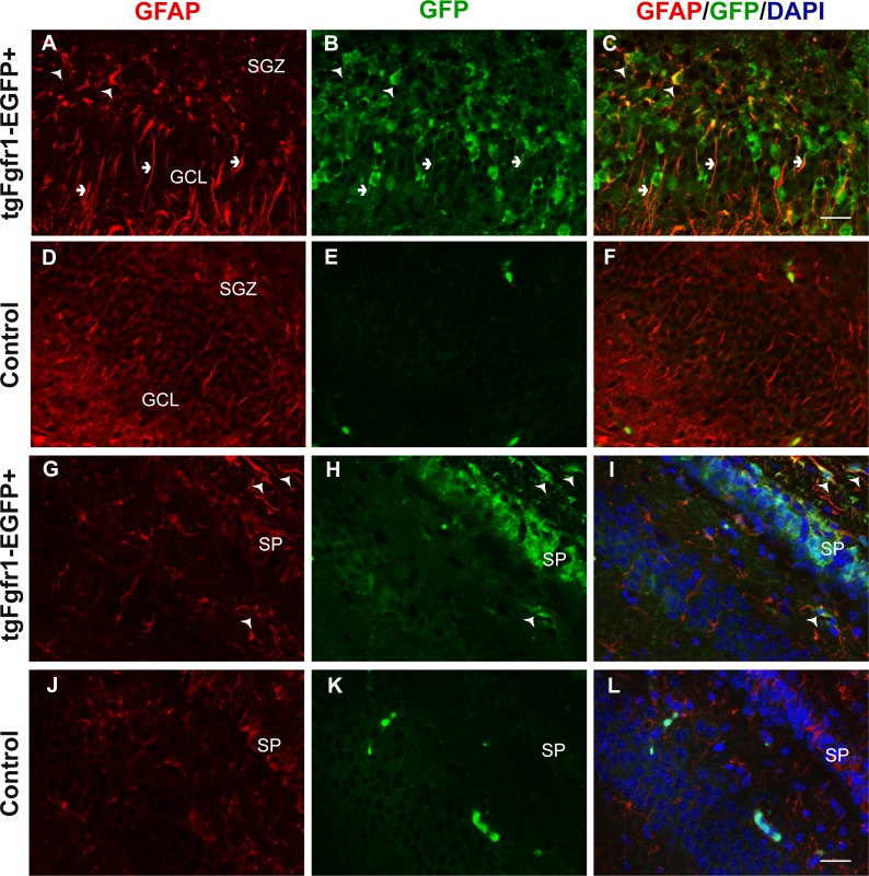
Figure 1. Fgfr1 expression in GFAP+ cells of the hippocampus at P7.
GFAP (A, D) GFP (B, E) immunostaining of the DG in P7 tgfgfr1-EGFP+ mice (A–C, n = 3) and tgfgfr1-EGFP-littermate controls (D–F, n = 3) demonstrated strong GFAP/GFP colocalization in cells of the SGZ (arrowheads) and their radial fibers into the GCL (small arrows). GFAP and GFP immunostaining of the CA region in tgfgfr1-EGFP+ mice (G–I, n = 3) and tgfgfr1-EGFP-controls (J–L, n = 3). GFP+ staining was observed in stratum pyramidale (SP) as well as in cells above (stratum oriens, SO) and below (stratum radiatum, SR) this layer (H). GFAP+/GFP+ colabeling (arrowheads) was observed in primarily in the SO and SR within the CA region, and in the white matter dorsal to the hippocampus. Scale bar is 25 µm. SGZ, subgranular zone; GCL, granule cell layer; SP, stratum pyramidale.

