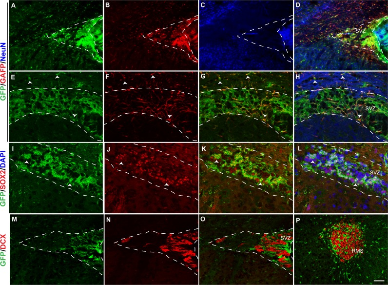
Figure 6. Fgfr1 expression in the SVZ and rostral migratory stream of 1-month mice.
GFP (A, E, D, H), GFAP (B, F, G, H), NeuN (C, D), immunostaining of the SVZ in 1-month tgfgfr1-EGFP+ mice (A–H, n = 3). GFP+ cells of the SVZ colocalized with GFAP+ cells (D, and G, H. Arrowheads in E–H = GFAP/GFP+ cells and GFAP+ fibers. GFP (I, K, L) and SOX2 (J, K, L) staining demonstrated that many, but not all SOX2+ cells also colabel with GFP+ (K and L, arrows indicate double labeled cells, DAPI staining included in L). In contrast, DCX+ neuroblasts in the SVZ and rostral migratory stream did not colabel with GFP (M–P). Scale bar is 50 µm in A–D and M–P and 25 µm in E–L. Dashed lines indicated SVZ region examined.

