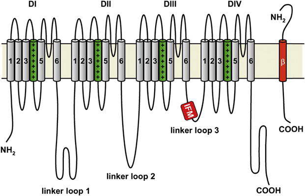
Figure 1. Membrane topology of NaV1.5.

Illustration of the four domains (DI–DIV) of the α subunit along with an associated β subunit. The transmembrane S1–S4 helices that comprise the voltage sensing domains are depicted with the charged S4 helix highlighted in green (dark gray in print versions). The S5 and S6 helices form the central pore domain. The DIII–DIV inactivation loop is highlighted with the critical hydrophobic isoleucine, phenylalanine, and methionine (IFM) sequence. The N-terminal and C-terminal domains of each subunit also labeled. Copyright Hugues Abriel, Oxford University Press, 2007. http://dx.doi.org/10.1016/j.cardiores.2007.07.019
