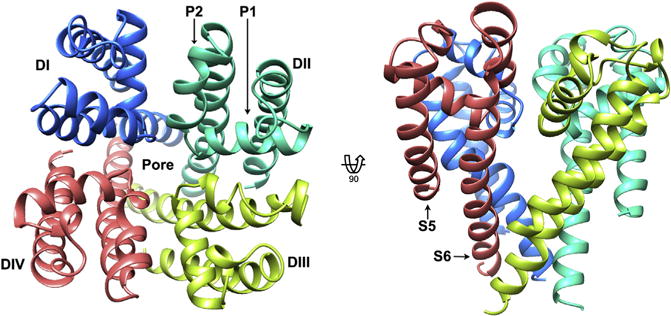
Figure 2. Homology model of the NaV1.5 channel pore domain.

Top view of the extracellular face (left), and transmembrane view (right), of the pore domain of NaV1.5, based on the bacterial template NaVRh (PDB ID code 4DXW). Structure is shown in ribbon representation with each domain colored from DI (blue (dark gray in print versions)) to DIV (red (light gray in print versions)). Pore helices P1 and P2 are labeled in DII, and transmembrane helices S5 and S6 are labeled in DIV for representative purposes. Kevin R. DeMarco.
