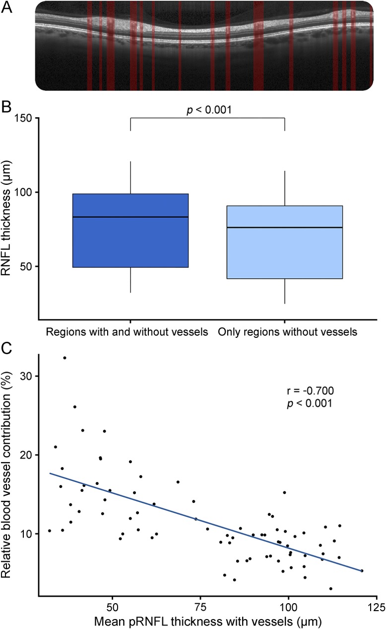
Figure. Contribution of blood vessels to retinal nerve fiber layer thickness.

(A) pRNFL scanned by optical coherence tomography, segmented with vessel detection. (B) Mean pRNFL thickness in μm; all regions including vessels and only regions without vessels. (C) Correlation of the relative blood vessel contribution to the pRNFL including vessels. pRNFL = peripapillary retinal nerve fiber layer.
