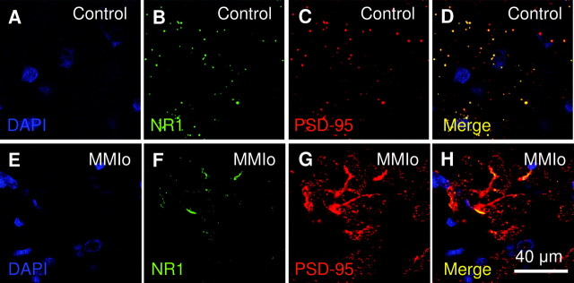
Fig. 6.
NR1 and PSD-95 localization in the hippocampus of rats gestated in hypothyroxinemic mothers. The NR1 and PSD-95 colocalization were analyzed in CA1 area of the hippocampus of MMIo or Co groups by confocal microscopy. A and E show DAPI staining (blue) for nucleus identification at the CA1 area. B and F, NR1 staining (FITC, green). C and G, PSD-95 staining (Cy3, red). D and H, The merger of the three fluorochromes DAPI, FITC, and Cy3, to analyze the colocalization of NR1 and PSD-95. Bar, 40 μm. A–D correspond to confocal microscopy pictures of the Co group. E–H correspond to confocal microscopy pictures of the MMIo group.

