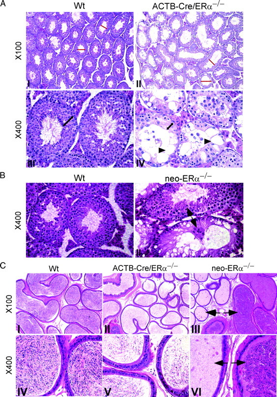
Fig. 3.

Histological analyses and comparison of testes and epididymides in the 12-wk-old Wt, neo-ERα−/−, and ACTB-Cre/ERα−/− males. A, Histological analyses of testes from Wt and ACTB-Cre/ERα−/− males. The adult Wt testes consist of compact seminiferous tubules (I and III). The adult ACTB-Cre/ERα−/− testes consist of atrophic and degenerating seminiferous tubules (red line) (II). The seminiferous epithelial layer in the ACTB-Cre/ERα−/− testes contained few spermatogenic cells with decreased germ cell number compared with Wt (IV vs. III, double-headed arrows), and the vacuoles were often present in the seminiferous epithelial layer (IV, arrowhead). B, Histological analyses of testes from Wt and neo-ERα−/− males. The adult neo-ERα−/− testes showed the defects with less extent compared with ACTB-Cre/ERα−/− testes. Some normal seminiferous tubules could be observed in neo-ERα−/− testes (II, arrow). C, Histological analyses of caudal epididymis from Wt, ACTB-Cre/ERα−/−, and neo-ERα−/− males. The adult Wt epididymis is full of sperm in the caudal lumen (I and IV). The caudal epididymis in the adult ACTB-Cre/ERα−/− males contains much less sperm and appears pale in the histology staining (II and V). The epididymis in the adult neo-ERα−/− males exhibits the heterogeneous phenotype. Some epididymal tubes contain very few sperm, but some are full of sperm (III and VI, arrows).
