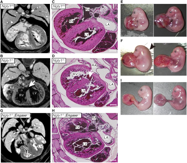
Fig 2. Loss of Ngly1 causes ventricular septal defects (VSD) and the additional Engase deletion rescues the VSD phenotypes.
(A, B, G) Maximum intensity projection of heart μ-CT images of E16.5 embryo of wild-type (A), Ngly1−/− (B), and Ngly1−/−;Engase−/− (G). White arrows in (B) indicates VSD. (C, D, H) Transverse section of wild-type (C), Ngly1−/− (D), and Ngly1−/−;Engase−/− embryo (H) at E16.5 were stained with H&E. White arrows in (D) indicate VSD. Shown are representative sections (n = 3). Scale bar in (C), (D) and (H) indicate 200 μm. RA: right atrium, RV: right ventricle, LA: left atrium, LV: left ventricle. (E) Anemia was observed in Ngly1−/− embryo at E16.5 (left panel). Right panel shows Ngly1+/+ embryo at E16.5 (littermate of the left panel). (F) Edema was observed in Ngly1−/− embryo at E16.5 (left panel). Right panel shows Ngly1+/+ embryo at E16.5 (littermate of the left panel). Black arrowhead indicates edema. (I) Anemia was observed in Ngly1−/−;Engase−/− embryo at E16.5 (left panel). Right panel shows Ngly1+/+;Engase−/− embryo at E16.5 (littermate of the left panel). Representative images were shown.

