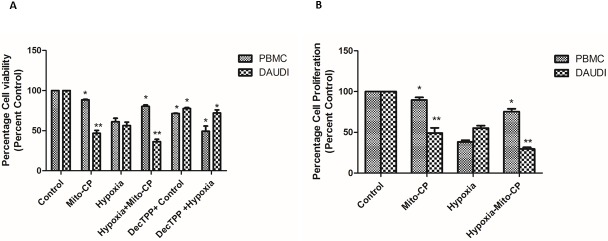
Fig 1. Effect of Mito-CP on cell viability and cell proliferation in Daudi cells and PBMCs by alamarBlue assay.
Daudi cells and PBMC were treated with and without Mito-CP under hypoxia (5% O2) and normoxia. (A) Shows percentage of cell viability after 6 h in Daudi and PBMC treated with and without Mito-CP and Dec-TPP+ under hypoxia and normoxia. Bar graph plotted represents percentage of viable cells normalised value to percent control. Data were obtained from three separate experiments and were expressed as by mean ± SEM. * and ** denotes significantly different compared to control p<0.05 and p<0.01 respectively. (B) Shows percentage of cell proliferation after 24 h in Daudi and PBMC treated with and without Mito-CP under hypoxia and normoxia. Each bar graph represented was normalised to percent control. Data were obtained from three separate experiments and are expressed as mean ± SEM. * and **, significantly different compared to control p<0.05 and p<0.01 respectively.

