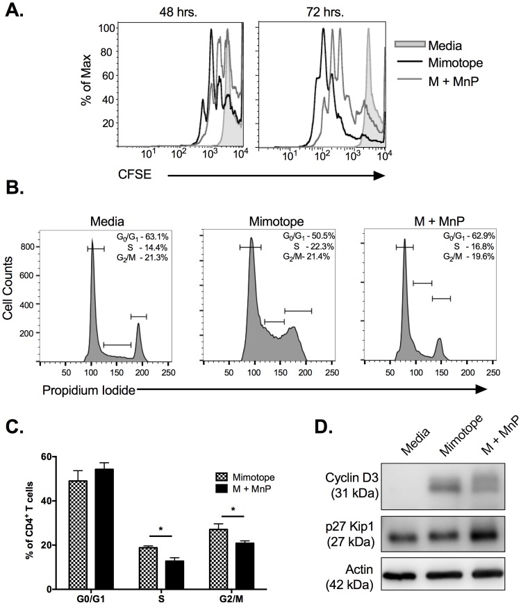
Fig 2. Redox modulation during activation inhibits cell cycle progression of CD4+ T cells.
NOD.BDC.2.5.TCR.Tg splenocytes were plated in complete splenocyte media and stimulated with 0.05 μM mimotope with or without 68 μM MnP (M + MnP) for 48–72 hours. (a) Prior to stimulation cells were loaded with 1 μM CFSE. Cells were stained with CD4 following harvest and analyzed by flow cytometry for CFSE dilution, indicating proliferation. Unstimulated cells (grey shaded curve) served as negative controls to set proliferation gates. CFSE tracings are representative of n = 6 independent experiments. (b) Cells were fixed, permeabilized, and stained with propidium iodide and CD4-FITC. Cells were analyzed by flow cytometry to determine cell cycle status. CD4+ T cells were gated on and cell cycle phases were set based upon unstimulated controls (left panel). (c) Percentages of n = 5 experiments were combined and graphed as mean ± SEM (* = p<0.05). (d) 48 hrs. post-stimulation, cells were harvested and analyzed by Western blot for p27 Kip1 and Cyclin D3 expression. β actin expression served as the loading control.

