
Fig 6.
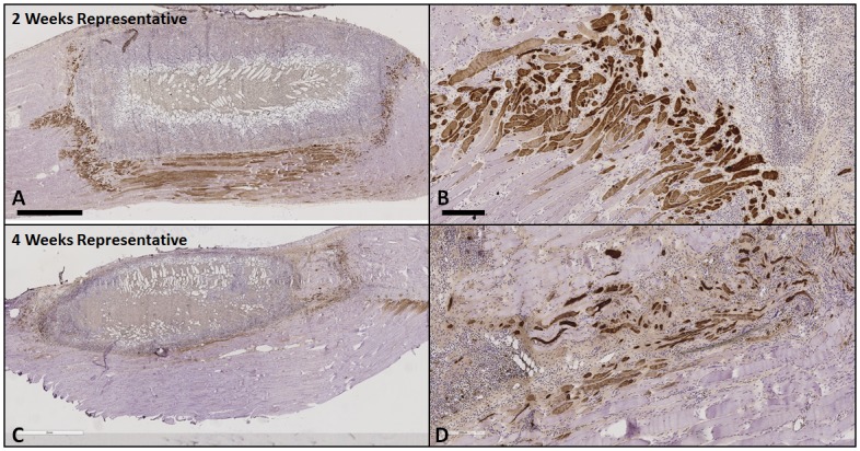
Represenative sections from a 2 week (A and B, gel only) and 4 week (C and D, gel only) TA muscle explant showed that embryonic myosin (f1.652+) was only observed in proximity to the edges but not within the defect, providing evidence for regenerating host muscle fibers that were injured during surgery but not neo-muscle formation in the defect. The density of f1.652+ fibers was observed to be higher at 2 weeks than 4 weeks. Scale bar = 2 mm in A and C. Scale bar = 200 μm in B and D.