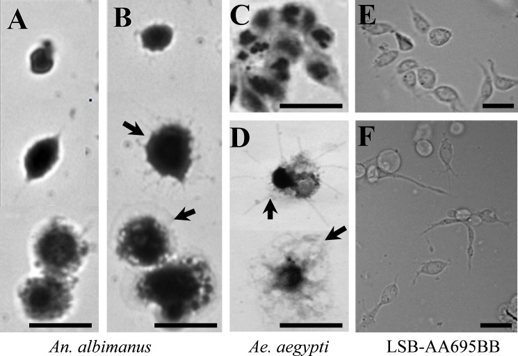
Fig 1. AT induced phenotypic changes on mosquito hemocytes and LSB-AA695BB cells.
Hemocytes from An. albimanus (A-B) and Ae. aegypti (C-D) were obtained by perfusion and incubated in a humid chamber with: Grace´s medium alone (A, C) or AT (10−7 M) (B, D). After 30 minutes, samples were fixed, stained with Giemsa and observed by bright-field microscopy. LSB-AA695BB cells were grown in a glass cover-slide, and then incubated 30 min in: Schneider´s medium alone (E) or containing AT (10−7 M) (F). Samples were observed by phase contrast microcopy. Hemocyte spreading (arrows). Scale bars: 5μm.

