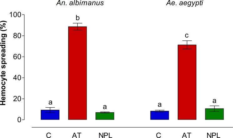
Fig 2. Hemocyte spreading percentages in An. albimanus and Ae. aegypti.
Mosquito hemocytes were obtained by perfusion, and incubated in a humid chamber with either Grace´s medium alone (C), AT (10−7 M) or NPLP1 (NPL, 10−7 M). After incubation, samples were analyzed by contrast phase microscopy and hemocytes showing spreading were recorded. Each hemocyte sample from groups of five mosquito per treatment was individually analyzed. Three independent experiments were performed. Data are expressed as percentage of spreading (Mean ± SEM). Significant differences were determined with one-way ANOVA followed by Tukey’s test (p < 0.001). Different letters on the top each bar are significantly different.

