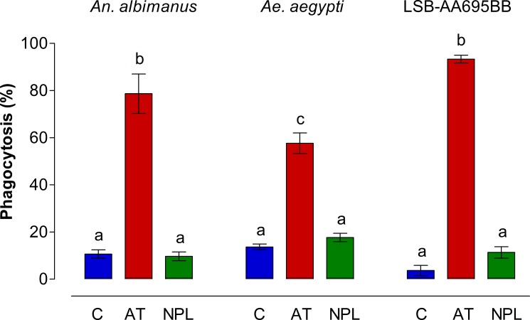
Fig 4. AT increases phagocytic activity by mosquito hemocytes and LSB-AA695BB cells.
Number of cells displaying fluorescent phagocytic vacuoles in hemocytes and LSB-AA695BB after incubation with medium (C) or with 10−7 M of Aedes-AT (AT) or NPLP1 (NPL). A minimum of 100 cells/sample (by triplicate) were counted for each treatment in three independent biological replicates. Data are expressed as percentage of phagocytic hemocytes (Mean ± SEM). Significant differences were determined with one-way ANOVA followed by Tukey’s test (p < 0.05). Different letter on the top each bar are significantly different.

