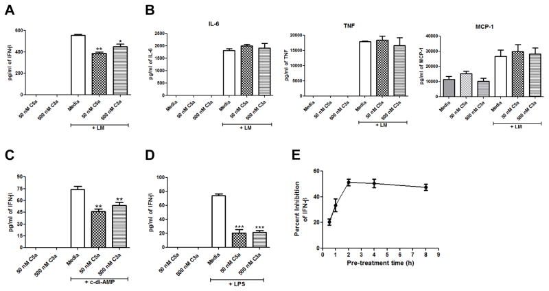
Figure 1. C5a and C3a suppress IFN-β production in J774A.1 cells.
J774A.1 cells were pre-treated with either media, 50 nM C5a, or 500 nM C3a for 2 h and then the cells were infected with Lm for 20 h. Cell-free supernatants were used to quantitate (A) IFN-β production or (B) IL-6, TNF-α, and MCP-1 production. J774A.1 cells were pre-treated with either media, 50 nM C5a, or 500 nM C3a for 2 h and then were incubated with (C) 25 μg/ml c-di-AMP or (D) 100 ng/ml LPS for 20 h. IFN-β was quantitated from cell-free supernatants. (E) J774A.1 cells were pre-treated with either media or 50 nM C5a for 30 min, 1 h, 2 h, 4 h, or 8 h and then were incubated with 100 ng/ml LPS for 20 h. IFN-β was quantitated from cell-free supernatants. All data are presented as mean pg/ml ± SEM. These data are pooled from three independent experiments. * P = 0.027; ** P ≤ 0.006; *** P ≤ 0.0001 by t test.

