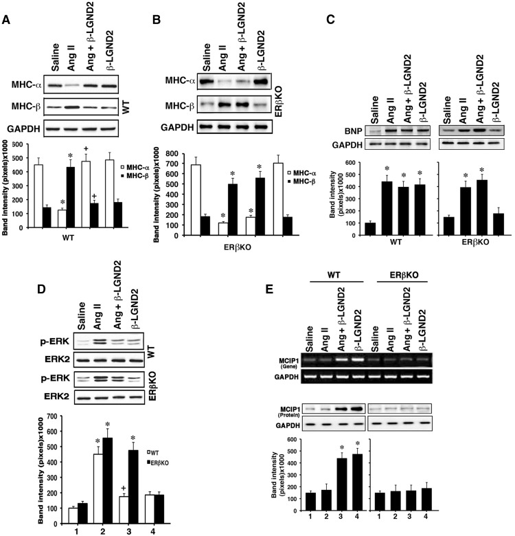
Figure 2.
E2 and an ERβ agonist modulates important markers of and signals for cardiac hypertrophy. A, MHC-α and -β proteins were determined by immune blot from the ventricles of each of six WT mice per condition. GAPDH is shown as a loading control, and blots are from a single representative mouse from each group. B, Same studies in ERβ KO mice. *, P < .05 comparing saline vs AngII infusion in WT mice and saline vs AngII or AngII + β-LGND2 in ERβ KO mice; +, P < .05 for AngII vs AngII + β-LGND2 in WT mice. C, Endogenous BNP protein in the left ventricles. Individual mice ventricles were processed with protein extraction for BNP expression by immune blot (n = 6 per condition). Single representative results from each condition are shown. *, P < .05 for saline compared with AngII or β-LGND2 infusion or both together in both mouse types. D, Activation of ERK by AngII is inhibited by β-LGND2 in WT mice. Kinase activity in the LV was determined at 3 weeks of treatment from equal amounts of ERK1 and ERK2 proteins immunoprecipitated from the mouse hearts and used for activating phosphorylation-immunoblots of ERK1 and ERK2. Total ERK2 protein is shown as loading control. Bar graph is the mean ± SEM densitometries from the samples (n = 6). *, P < .05 for control vs AngII in both models or AngII + β-LGND2 in ERβ KO mice; +, P < .05 for AngII vs AngII+ β-LGND2 in WT mice. E, MCIP1 mRNA (top panel) and protein (bottom panel) are stimulated by β-LGND2 in WT mice ventricles. mRNA expression was determined by RT-PCR, and protein was demonstrated by immunoblots with representative single samples per each condition shown. Bar graph of the mean ± SEM densitometry protein data are from individual mouse results combined. *, P < .05 for saline control vs β-LGND2 or β-LGND2 + AngII in WT mice.

