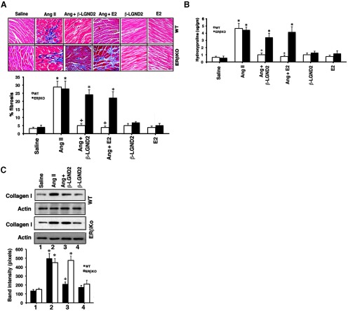
Figure 4.
β-LGND2 prevents AngII-induced cardiac fibrosis. A, Representative Masson trichrome staining of collagen deposition in the LV of ovariectomized female mice exposed to the indicated conditions is shown (n = 5 mice per condition). Arrows indicate fibrosis. Bar, 0.1 mm. The percent area of fibrosis is quantified as described in Materials and Methods, and the bar graph data are the mean ± SEM (n = 5 mice per condition). *, P < .05 for saline control vs AngII in either mouse type or versus β-LGND2 or E2 + AngII in ERβKO mice. +, P < .05 for AngII vs AngII + β-LGND2 or E2 in WT mice. B, Hydroxyproline content of the ventricle was measured by spectrophotometry and mean ± SEM was calculated from individual data from each ventricle. *, P < .05 for saline control vs AngII in either mouse type or vs β-LGND2 or E2 + AngII in ERβ KO mice; +, P < .05 for AngII vs AngII + β-LGND2 or E2 in WT mice. C, Collagen I protein expression is inhibited by estrogenic compounds in WT mice. Actin serves as the protein loading control, and bar graphs are mean ± SEM. *, P < .05 for saline vs AngII; +, P < .05 for AngII vs the same + β-LGND2 or E2 in WT mice.

