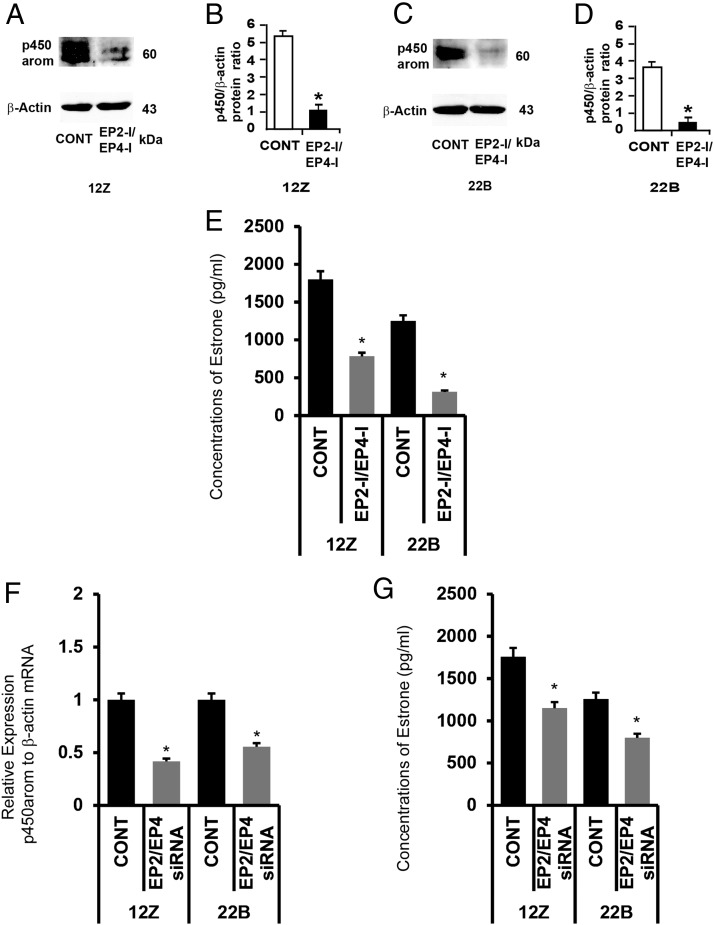
Figure 7.
Effects of inhibition of EP2 and EP4 receptors on activity of P450 aromatase (P450 arom) in human endometriotic epithelial cells 12Z and stromal cells 22B. (A and B) Western blot analysis of P450 arom in 12Z and 22B cells. β-Actin protein was measured as an internal control. (C and D) Densitometry for P450 arom performed by Alpha Imager and expressed in integrated density value (IDV). The 12Z and 22B cells were treated with inhibitors for EP2 (AH6809, 75μM) and EP4 (AH23848, 50μM) for 24 hours. Numerical data are expressed in ratio between P450 arom protein and β-actin protein as mean ± SEM of 3 (n = 3) independent experiments. CONT, control; *, CONT vs CTZ on expression of P450 arom protein in 12Z and 22B cells, P < .05. (E) Concentrations of estrone. The 12Z and 22B cells were treated with inhibitors (EP-I) for EP2 (AH6809, 75μM) and EP4 (AH23848, 50μM) for 24 hours. The culture media were collected, and concentration of estrone was measured using ELISA. Numerical data are expressed as mean ± SEM of 3 (n = 3) independent experiments. *, CONT vs EP2-I + EP4-I on estrone biosynthesis by 12Z and 22B cells, P < .05. (F and G) Effects of knock down of EP2 and EP4 genes P450 arom gene expression and estrone production. (F) Expression of P450 arom mRNA was performed using qPCR, and data were analyzed using dCT method. (G) Concentration of estrone in the culture media were measured using ELISA. Numerical data are expressed as mean ± SEM of 3 (n = 3) independent experiments. More details are provided in Materials and Methods and Result sections.

