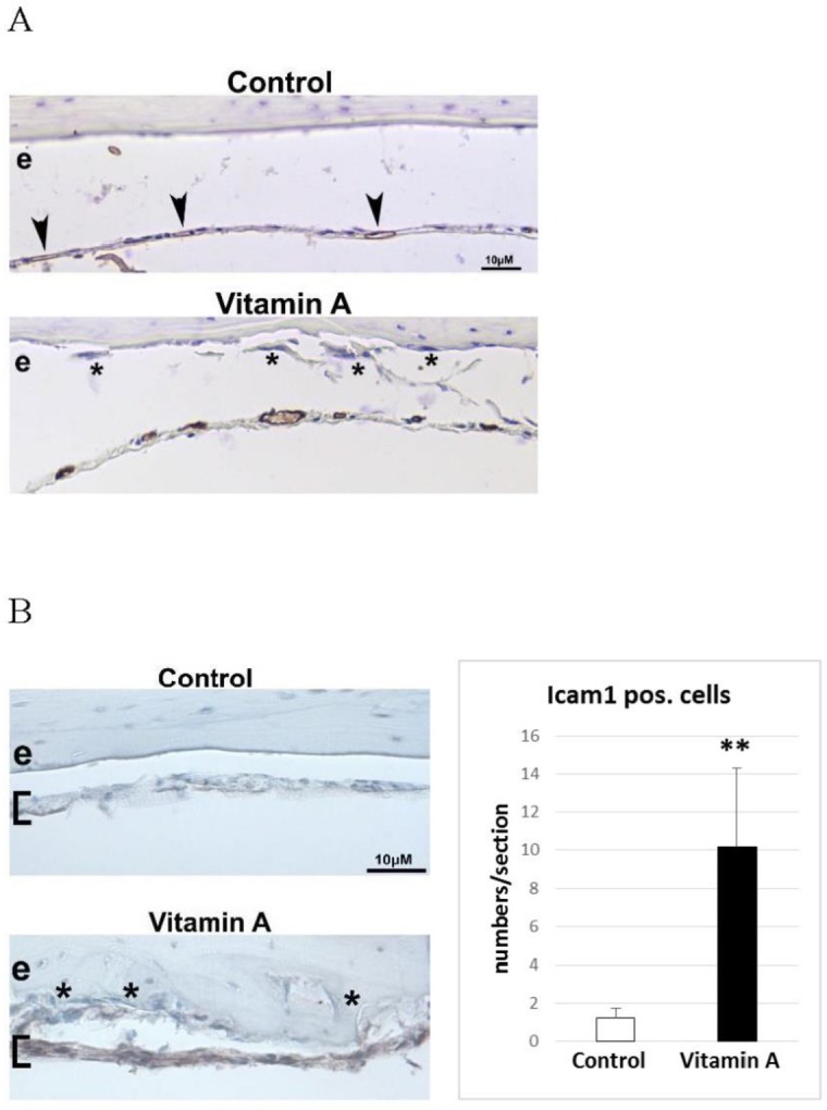
Fig 3. Endocranial associated dura mater blood vessels and Icam1 positive cells.

A) Representative immunohistochemical staining (brown) for platelet/endothelial cell adhesion molecule 1 (Pecam1) positive blood vessels in decalcified calvaria sections. Arrowheads indicate small and flat Pecam1 positive blood vessel present in the dura mater membrane (covering the endocranial bone surface) of control tissue. In the “Vitamin A” panel the Pecam1 positive blood vessels are readily visible and appear engorged. Asterisks indicate osteoclasts in close proximity to the enlarged vessels, a combination only found in vitamin A animals. e = endocranial bone surface B) Immunohistochemical staining (brown) for intercellular adhesion molecule 1 (Icam1) in decalcified calvaria sections. Brackets indicate position of the dura mater membrane which is highly Icam1 positive only in vitamin A animals. Asterisks indicate osteoclasts in close proximity to the Icam1 positive cells, only found in vitamin A animals. Right panel shows the number of Icam1 positive cells/section in the dura mater (n = 4 and 6 sections/group and each group contain sections from 3 different individuals). Results are given as means + SD. ** p < 0.01.
