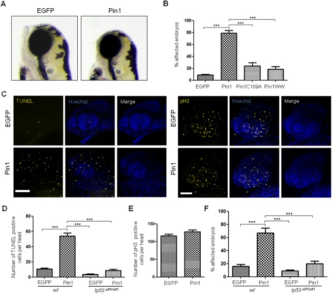
Fig 5. pin1 mRNA injection affects head development in zebrafish.
(A) Lateral views of live 3 dpf embryos injected with EGFP mRNA or EGFP-Pin1 mRNA (Pin1) at 1–2 cell stage. (B) Quantification of the percentage of embryos with altered head development upon injection of mRNAs coding for EGFP, EGFP-Pin1, EGFP-Pin1C109A or EGFP-WW, as indicated. Embryos with evident facial retraction, reduced mandible and/or reduced head size were scored as positive. The graph shows the average of 4 experiments with at least 50 embryos each and the standard deviation. Statistical analysis were carried out using one-way ANOVA followed by Tukey's Multiple Comparison Test: *** p˂ 0,0001. (C) Representative confocal projections of embryos microinjected with EGFP mRNA or EGFP-Pin1 mRNA, stained for apoptosis by whole-mount TUNEL assay (left panels) or for proliferation by p-H3 whole-mount immunofluorescence (right panels). (D) Quantification of apoptotic cells on heads of wild type (wt) or tp53 zdf1/zdf1 embryos microinjected with EGFP mRNA or EGFP-Pin1 mRNA as indicated. Statistical analyses were carried out using one-way ANOVA followed by Tukey's Multiple Comparison Test: *** p˂ 0,0001 (n = 17 for each condition). (E) Quantification of cells positive for p-H3 staining on heads of wild type embryos microinjected with EGFP mRNA or EGFP-Pin1 mRNA as indicated. Statistical analyses were carried out using unpaired t test with Welch's correction (n = 21 for each condition). (F) Morphological analysis at 3 dpf of wild type or tp53 zdf1/zdf1 embryos microinjected with EGFP mRNA or EGFP-Pin1 mRNA as indicated. The graph shows the average of 4 experiments with at least 50 embryos each and the standard deviation. Statistical analysis were carried out using one-way ANOVA followed by Tukey's Multiple Comparison Test: *** p˂ 0,0001.

