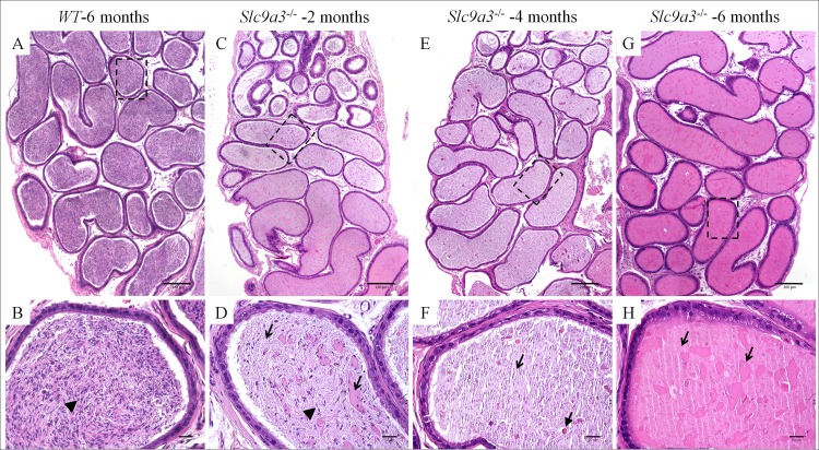
Fig 7. Slc9a3-/- cauda epididymis is filled with abundant abnormal secretions instead of spermatozoa.
H&E-stained sections of the cauda epididymis of 6 month-old WT (A, B) and Slc9a3–/–males aged 2 months (C, D), 4 months (E, F), and 6 months (G, H). (A, C, E, G) The low magnification overview is a montage assembled by an array of films continuously taken with a ×10 objective on a bright microscope. (B, D, F, H) Magnified view of the area indicated by a dashed box in the upper panel. Spermatozoa are indicated by arrowheads, and abnormal heterogeneous secretions are marked by arrows, a dashed arrow, and a green arrow. The white lines are sectioning artefacts. Scale bar = 200 μm (A, C, E, G) and 20 μm (B, D, F, H).

