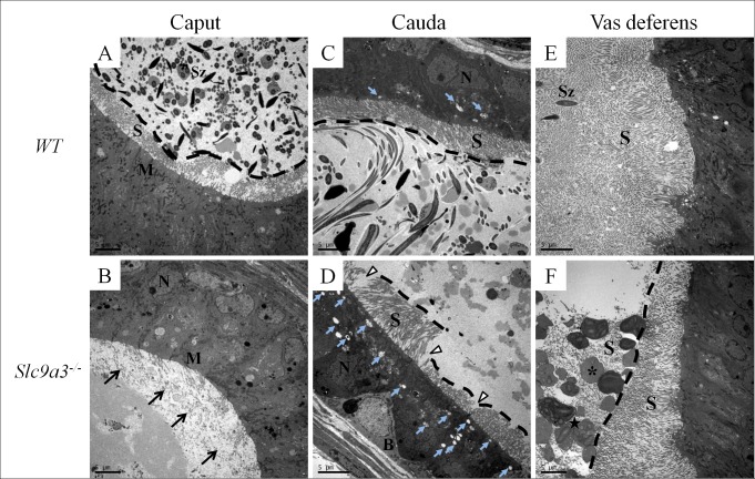
Fig 9. Ultrastructural defects of the epithelium in Slc9a3–/–epididymis and vas deferens.
An electron micrograph (cross-section) showing the ultrastructure of the epithelia of the excurrent ducts of 2-month-old WT (upper panel) and Slc9a3-/- male mice (lower panel). (A, B) Caput epididymis. The arrow indicates the absence of stereocilia on the caput epithelium. The black dashed line marks the boundary between the lumen and the edge of stereocilia on the epithelium. (C, D) Cauda epididymis. The white arrowhead indicates the disturbed and decreased number of stereocilia. The light blue arrow indicates the vesicles in the cauda epithelium. (E, F) Vas deferens. The asterisk indicates abnormal secretory particles and the star indicates smooth endoplasmic reticulum-like material in the particles (enlarged image in S2 Fig). The magnification for each image is slightly different, with 2000× magnification used for A, B, and E and 2500× magnification used for C, D, and F. Scale bar = 5 μm. Abbreviations: B, basal cell; M, mitochondria; N, nucleus; S, stereocilia; Sz, spermatozoa.

