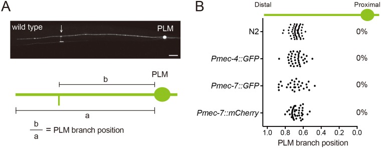
Fig 1. The PLM branching positions are highly predictable.
(A) Schematic diagram of PLM morphology. (B) PLM branching pattern identified by immunostaining of N2 wild type with K40-acetylated tubulin antibody or by the transgenes zdIs5(Pmec-4::GFP), jsIs973(Pmec-7::RFP) and muIs42(Pmec-7::GFP). The percentage of mislocalized PLM branching sites was shown at the right of the distribution plot. N > 25.

