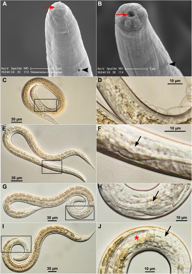
Fig 1. Morphological changes of activated S. carpocapsae nematodes.
(A) The head region of a non-activated IJ by scanning electron microscopy (SEM). (B) The head region of an activated nematode by SEM. The mouth is marked by a red arrow and the excretory pore by a black arrowhead in (A) and (B). (C) A non-activated IJ by light microscopy (400x). (D) An enlarged view of the boxed region in (C). (E) A partially activated nematode (400x). (F) An enlarged view of the boxed region in (E) where the partially expanded terminal pharyngeal bulb (arrow) is located. (G) A fully activated nematode (400x). (H) An enlarged view of the boxed region in (G). (I) A fully activated nematode whose gut is wide open (400x). (J) An enlarged view of the boxed region in (I). The black arrowhead in (A) and (B) point to the secretory/excretory pore. The black arrows in (F), (H) and (J) point to the pharyngeal bulbs. The red star in (J) marks the open gut.

