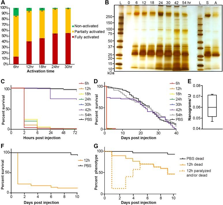
Fig 2. Activated S. carpocapsae ESPs contain lethal proteins.
(A) In vitro activation rates of IJs over a time course. Error bars represent standard errors. (B) A silver-stained protein gel of ESPs from nematodes that have been activated for different lengths of time, and ESPs from axenic nematodes. The left-hand side of the gel shows the secreted proteins from symbiont-associated IJs activated for different amounts of time. The right-hand side of the gel shows the proteins secreted from symbiont-associated (S) and axenic (A) IJs that were exposed to waxworm homogenate for 12 h. Each lane contains 1% of the total ESP volume. The 0h sample was collected from non-activated IJs. L, protein ladder; S, symbiotic IJs; A, axenic IJs. (C) Survival curves within 72 hrs of Drosophila injected with 20 ng of ESPs from nematodes having been activated for different lengths of time. The 30 hr curve (blue) mostly overlaps with the 6 hr curve (red). (D) Survival curves over 40 days of Drosophila injected with 20 ng of ESPs from nematodes having been activated for different lengths of time. (E) The average amount of ESPs secreted by individual IJs in 3 hours. (F) Survival curves of 2nd instar Bombyx mori larvae injected with 650 ng of ESPs from axenic nematodes that were activated for 12 h. (G) Phenotype curves of last instar Galleria mellonella larvae injected with 4 μg of ESPs from axenic nematodes that were activated for 12 h. The dotted line shows the number of larvae that were either killed or paralyzed after the injection. Paralyzed waxworms recovered over time.

