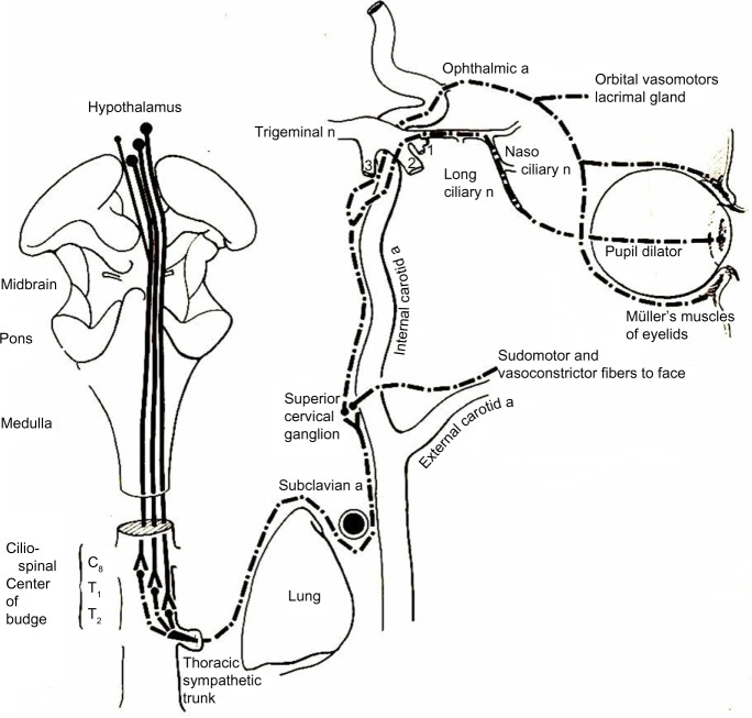
Figure 2.
Drawing showing the anatomy of the oculosympathetic pathway.
Notes: Sympathetic fibers in the posterolateral hypothalamus pass through the lateral brain stem and to the ciliospinal center of Budge and Waller in the intermediolateral gray column of the spinal cord at C8–T1. Preganglionic sympathetic neurons exit from the ciliospinal center of Budge and Waller and pass across the pulmonary apex and ascend up the carotid sheath to the superior cervical ganglion. The postganglionic sympathetic neurons originate in the superior cervical ganglion and travel up the wall of the internal carotid artery. Once the fibers reach the cavernous sinus, they travel with the abducens nerve before joining the ophthalmic division of the trigeminal nerve and entering the orbit with its nasociliary branch. From here, they divide into two long ciliary nerves to reach the iris dilator muscle. Copyright © 1978 Wolters Kluwer. Reproduced with permission from. Glaser JS, editor. Neuro-ophthalmology. 1st ed. Hagerstown MD, USA: Harper & Row; 1978.133
Abbreviations: a, artery; n, nerve.

