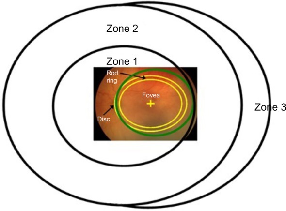
Figure 5.

Diagram of the International Classification of Retinopathy of Prematurity zones24 with a superimposed fundus photograph on which the optic disc and fovea are indicated.
Notes: The green circle indicates the region of retina viewed through a 28 diopter lens used with the indirect ophthalmoscope. The band delineated by the yellow lines represents the location of the anatomical “rod ring”,9 which is an annular region in which there is a high density of rods. The ring is concentric with the fovea and passes just nasal to the optic disc and approximately 18° temporal to the fovea.
