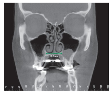
Figure 24. Coronal slice after expansion shows more favorable buccolingual inclination (torque) of posterior maxillary teeth. Also, nasal cavity floor is 23.2 mm wide, larger than at baseline (15 mm). Wax-bite registration was sent to the radiologic laboratory, but the CBCT scan was obtained at maximal intercuspation, suggesting that posterior left crossbite is still present. There was no reason to irradiate the patient again.