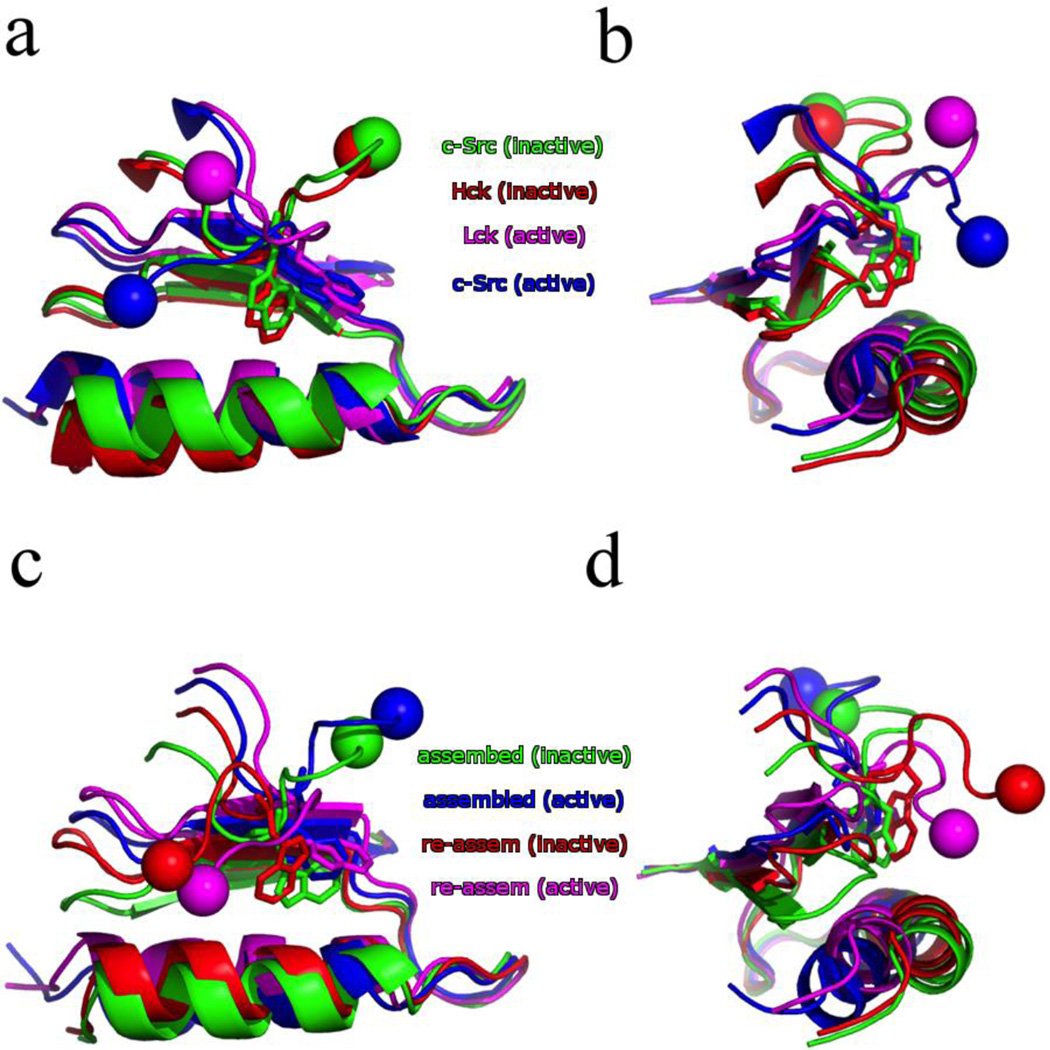
Figure 4.
Comparison of the W260 position in both the X-ray crystal structures (a–b) and converged string endpoints (c–d). The αC-helix and portions of the N-lobe are shown as well. The spheres indicate the direction of the N-terminal linker relative to the KD. The X-ray crystal structures shown are the inactive Hck (red, PDB id: 1QCF), inactive c-Src (green, PDB id: 2SRC), active Lck (magenta, PDB id: 3LCK), and active c-Src (blue, PDB id: 1Y57). The converged string endpoints are the assembled SH2/SH3 inactive (green) and active (blue), followed by the re-assembled SH2/SH3 inactive (red) and active (magenta).

