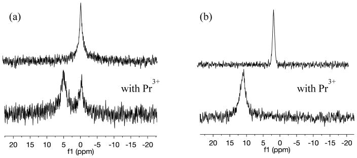
Figure 1.
Representative 31P NMR spectra of (a) 30 mM DMPC vesicles and (b) 30 mM DMPC and 20 mol% 6-kc (upper) and with addition of PrCl3.6H2O (lower). In pure DMPC vesicles, Pr3+splits the phosphorus signal into two, representative of inner and outer leaflets, while in vesicles containing 6-kc, it shifts the entire signal downfield indicating that it has permeated the bilayer.

