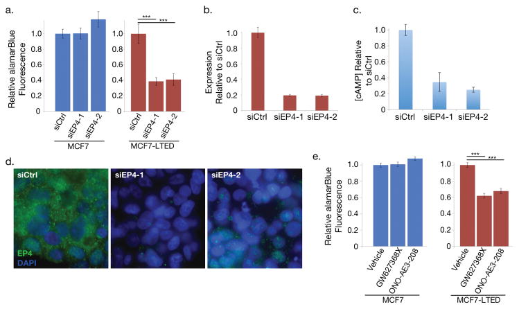
Figure 2. Knockdown or inhibition of EP4 signaling decreases estrogen independent cell proliferation.
(a) Proliferation of MCF7 cells, which express little EP4, treated with EP4 siRNA and MCF7-LTED cells relative to cells treated with negative control siRNA. (b) RT-qPCR analysis of PTGER4 expression decreases in MCF7-LTED cells treated with two distinct EP4 siRNAs relative to control siRNA. Error bars are standard deviation for three technical replicates. (c) cAMP levels of MCF7-LTED cells treated with EP4 agonist decrease in cells treated with siRNAs to EP4 relative to siRNA controls. (d) Immunofluorescence images of EP4 (green) and DAPI (blue) in MCF7-LTED cells treated with siRNAs targeting EP4 and siRNA controls. (e) Cell proliferation of EP4 antagonists in MCF7 and MCF7-LTED cells relative to cells treated with vehicle only. *** indicates p < 0.001. Error bars show s.e.m. of three replicates.

