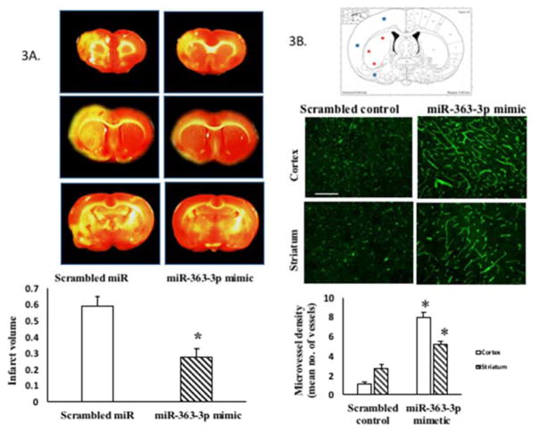
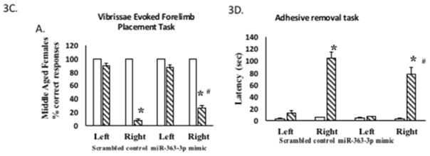
Figure 3. Effect of intravenous post stroke miR-363-3p mimic treatment to middle aged females.
(A) Infarct volume: TTC stained brain sections at 5d post stroke from middle-aged females treated with scrambled oligos (control) or miR363-3p mimic. Histogram depicts average infarct volume (±SEM) normalized to the volume of the non-ischemic hemisphere. B. Microvessel density: Schematic of a brain coronal section (bregma −4.0mm; Paxinos) depicts the three regions of the cortex and striatum that were photographed for microvessel density analysis. Photomicrograph of tomato lectin stained microvessels in the cortex and striatum of mir363-3p or control treated animals after MCAo. Histogram depicts mean (+SEM) density of Tomato lectin labeled microvessels. *: p<0.05. Bar=200 μm. C. Sensory motor performance was evaluated by vibrissae evoked forelimb placement task. Histogram depicts percent (+SEM) correct responses over 10 trials. D. Sensory motor performance on the adhesive removal test was evaluated before and after stroke. Histograms depict mean (±SEM) latency in seconds to remove the tape. (*: p<0.05, comparison of pre versus post stroke performance; #: p<0.05, control versus mir363-3p post stroke performance). In C and D, clear bars represent pre-stroke performance and hatched bars represent post stroke performance


