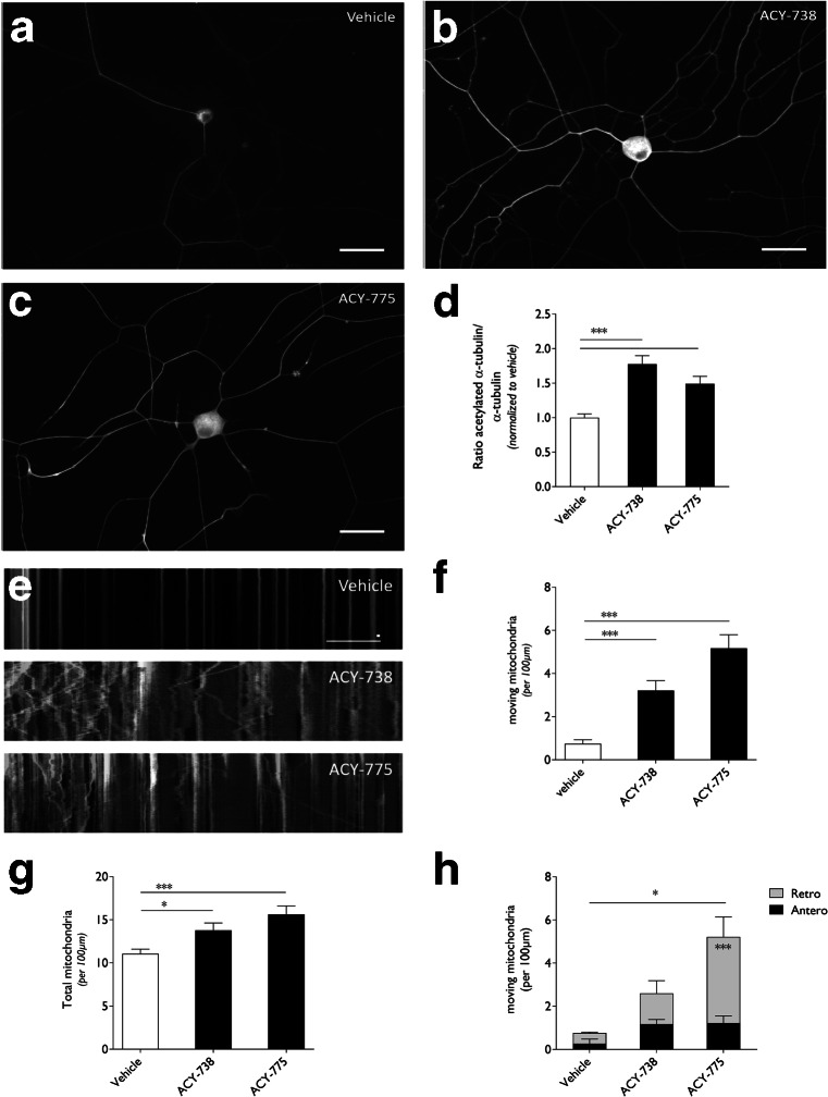
Fig. 3.
Histone deacetylase 6 (HDAC6) inhibition using ACY-738 and ACY-775 rescued the mitochondrial defects in dorsal root ganglion (DRG) neurons cultured from symptomatic HSPB1S135F mice. (a–c) Nontransgenic DRG neurons were incubated with 2.5 μM ACY-738 or ACY-775. Immunocytochemical labeling of acetylated α-tubulin shows increased immunofluorescence in neurons upon HDAC6 inhibition. Scale bar = 40 μm. (d) The intensity of acetylated α-tubulin was quantified in the neurites from DRG neuron cultures and corrected for the length of the signal. All values within each experiment were normalized to vehicle-treated cells; n = 5 with 43–61 cells per condition. (e) Kymographs representing the axonal movement of mitochondria in HSPB1S135F DRG neurons treated with ACY-738 or ACY-775 (2.5 μM). Vertical lines representing stationary mitochondria and lines deflecting to the right or left indicate anterograde or retrograde movement of mitochondria, respectively. (f) The number of moving mitochondria per 100 μm in the neurites was quantified based on the kymographs from HSPB1S135F DRG neurons treated with ACY-738 or ACY-775; n = 25–35 from 3 different transgenic mice. (g) The total number of mitochondria per 100 μm in the neurites was quantified based on the kymographs from HSPB1S135F DRG neurons treated with ACY-738 or ACY-775; n = 25–35 from 3 different transgenic mice. (h) Additional analysis was performed to assess the number of anterogradely or retrogradely moving mitochondria per 100 μm in the neurites based on the kymographs from HSPB1S135F DRG neurons treated with ACY-738 or ACY-775; n = 3 with 7–10 cells per experiment. One-way analysis of variance (ANOVA); *p < 0.05, ***p < 0.0001. Two-way ANOVA; *p < 0.05, ***p < 0.0001

