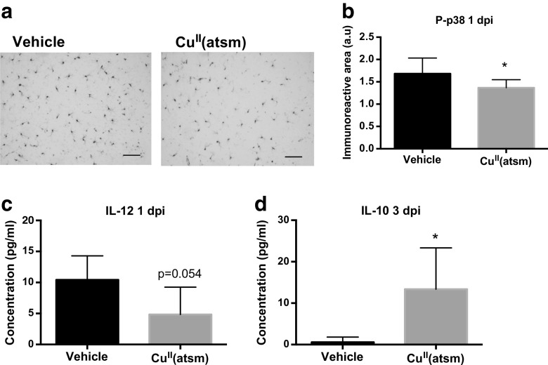
Fig. 7.
Copper delivery by CuII(atsm) reduces markers of inflammation in the ischemic brain. a Typical example images of phosphorylated p38 MAPK immunoreactivity in the peri-ischemic brain area in vehicle-treated and CuII(atsm)-treated mouse brains at 1 day after pMCAO. Scale bar = 20 μm. b Quantitative analysis of phosphorylated p38 MAPK immunoreactivity in the peri-ischemic brain area at 1 dpi. Data are shown as means ± standard deviation (SD). Unpaired 2-tailed t-test, *p < 0.05. n = 8 in both groups. c The concentration of IL-12 in the peri-ischemic brain area was measured by CBA at 1 dpi. Data are shown as means ± SD. Unpaired 2-tailed t-test, *p < 0.05. n = 5 in vehicle and n = 6 in the CuII(atsm) group. d The concentration of IL-10 in the peri-ischemic brain area was measured by CBA at 3 dpi. Data are shown as means ± SD. Unpaired 2-tailed t-test, *p < 0.05. n = 5 in vehicle and n = 6 in the CuII(atsm) group

